Image Based Gating of Intravascular Ultrasound Pullback Sequences
- Abstract
Intravascular Ultrasound(IVUS) is an imaging technology which provides cross-sectional images of internal coronary vessel structures. The IVUS frames are acquired by pulling the catheter back with a motor running at a constant speed. However, during the pullback, some artifacts occur due to the beating heart. These artifacts cause inaccurate measurements for total vessel and lumen volume and limitation for further processing. Elimination of these artifacts are possible with an ECG (electrocardiogram) signal, which requires a special gating unit and causes loss of important information about the vessel. To address this problem, we propose an image-based gating technique based on manifold learning and a novel weighted ultrasound similarity measure. Quantitative tests are performed on 12 different patients, 25 different pullbacks and 100 different longitudinal vessel cuts. In order to validate our method, the results of our method are compared to those of ECG-Gating method. In addition, comparison studies against the results obtained from the state of the art methods available in the literature were carried out to demonstrate the effectiveness of the proposed method.
- Gating Idea
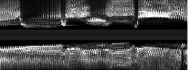
Longitudinal cut view of a nongated IVUS pullback shows a jagged character.
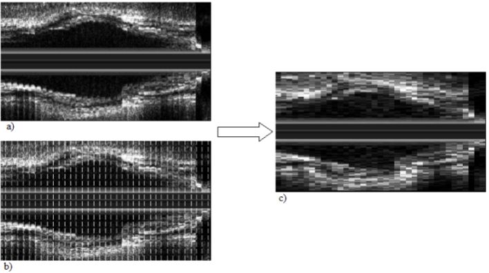
In this work, we introduce a robust image-based gating method based on manifold learning. By designing this method, our overall aim is to retain only the necessary information about the vessel, (the frames at a particular fraction of the RR-interval), which will adequately provide accurate lumen and vessel volume measurements; at the same time will avoid loss of important plaque information in the lesion areas.
Illustration of gating idea. a) Ungated pullback b)Dashed lines: The frames acquired in the same cardiac phase, c) Reconstruction of the pullback.
- Manifold Learning for Gating
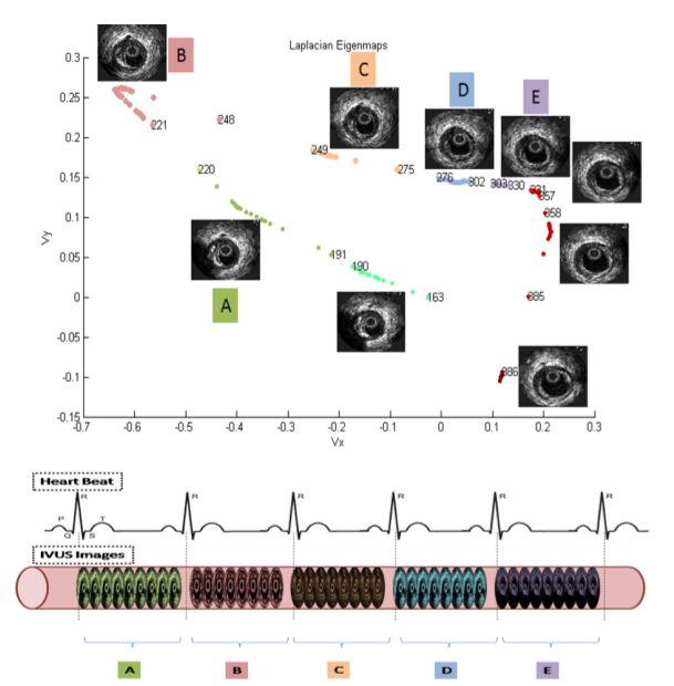
In order to illustrate the idea of manifold learning on our problem, a desired mapping for 9 sequential cardiac cycles is given in the figure below, where each IVUS image is projected onto a 2-D space that indicates a clustering among images. Each cluster is shown with a different color and assumed to represent a different cardiac cycle. Different cardiac cycles seem to be well-seperated from each other.
An illustration of Manifold idea. Each frame is shown with a dot on the calculated low-D manifold(here m=2), where A,B,C,D,E are the clusters of frames that belong to different cardiac cycles.
- Sample Result
First row: Nongated pullback. Middle Row; Left: Image-based gated pullback. Right: Ecg gated pullback. Bottom Row; Left: Manual lumen border of image based gated pullback. Right: Manual lumen border of ecg gated pullback.
- Acknowledgements
This project was developed with Matlab in 2010/2011 by Gozde Gul Isguder (Sabanci University- Masters Student) in collaboration with TUM. This project is financially supported by Tubitak (via Ass. Prof. Dr. Gozde Unal). Please see our publication list for further details or e-mail me.


